Uncovering the Past: Insights from an Ancient Skeleton
Written on
Chapter 1: Discovery of the Skeleton
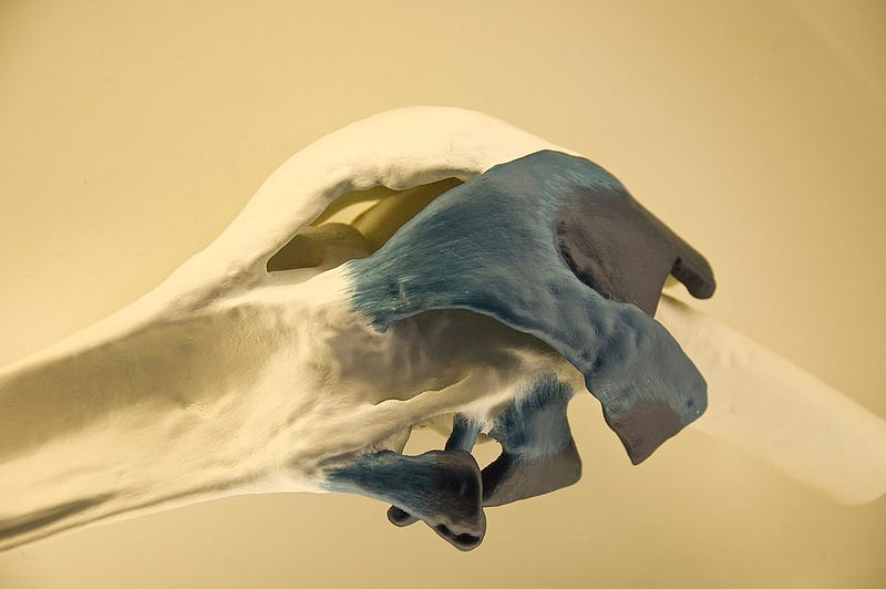
Archaeologists have unearthed the remains of a woman from several centuries ago in Portugal. This finding has revealed significant details about her health, particularly a notable bone growth on her femur. This abnormality is attributed to extraskeletal ossification, a condition that can occur following severe injury.

Researchers from the University of Lisbon investigated a female skeleton found in Constância, a town in central Portugal. The skeleton was located in a necropolis on the grounds of São Julião Church, alongside 151 other burials. It is believed that these remains date back to between the 14th and 19th centuries AD. Among their findings, the team noted a significant growth of nearly 8 centimeters on the upper part of the woman’s right thigh, indicative of extraskeletal ossification.
Section 1.1: Understanding Extraskeletal Ossification
The analysis determined that the bone growth was a result of extraskeletal ossification, a condition first documented in 1883. Following World War I, many soldiers returning from combat exhibited similar symptoms due to injuries to their musculoskeletal systems. This condition manifests as the formation of bone tissue in soft areas outside the skeletal structure.
“I have never seen such extensive ossification. This case piqued our interest. The femur's condition suggests a prolonged process of change,” explains Sandra Assis, an anthropologist at the University of Lisbon and the study's lead author. “While we lack the woman’s medical records, comparisons with similar clinical instances indicate that the femoral lesion was likely quite debilitating,” Assis elaborates.
Subsection 1.1.1: The Impact of Bone Growth
The ossification occurred at the junction of the muscle connecting the femur to the pubic bone, implying that the woman may have endured significant pain and limited mobility. The findings, published in the International Journal of Paleopathology, suggest that the lesion could stem from a single traumatic event or multiple injuries sustained within a short timeframe. The woman was approximately 50 years old at the time of her demise, and the lesion has been categorized as myositis ossificans traumatica, a form of traumatic ossification.
“This condition is frequently seen in younger males, particularly athletes, but historical records show cases across various ages and genders,” the researchers note.
Chapter 2: The Woman's Life and Labor
The woman’s skeleton, although incomplete, was remarkably preserved. The researchers estimated her height to be around 154 cm and concluded that the bone growth likely developed about a year before her death. They highlighted that the ossification drastically altered her quality of life.
“Today, such growths can be surgically removed, but during her lifetime, such medical intervention wasn’t an option,” Sandra Assis emphasizes.
The circumstances surrounding the woman’s occupation remain a topic of speculation. Constância has a historical reputation for its port and economy based on the Tagus River. In the 19th century, while women primarily engaged in domestic duties, many also worked as weavers or seamstresses.
At that time, heavy labor, often leading to serious injuries, was predominantly a male endeavor. However, the researchers propose that this woman may have been an anomaly, possibly working alongside men in tasks such as loading goods. An unfortunate accident could have led to the injuries that resulted in her condition.
Closure of Pyramid of Cheops: What it Means for Egyptian Tourism
One of the world’s most iconic tourist attractions, the Pyramid of Cheops in Giza, Egypt, is set to close soon…
Thank you for taking the time to read this article. If you found it valuable, please consider showing your appreciation by leaving some claps or following me. Your support is greatly appreciated!